Robert Peel
Guest

Patient Chart
| Patient Name: | Robert Peel | Date of Birth: | 04/03/1973 | Gender: | Male | ||||
| Address: | 10 Downing Street | Allergies: | NKDA | Presenting Complaint: | Trauma | ||||
Vitals | GP: | West London Clinic | |||||||
| Time: | 08:00 | 08:05 | 08:10 | 08:15 | 08:20 | 08:25 | 08:30 | 08:35 | 08:40 |
| Heart Rate: | 110 | 115 | 101 | 105 | 110 | 115 | 115 | 120 | 125 |
| Respiratory Rate: | 24 | 26 | 24 | 22 | 24 | 20 | 20 | 18 | 18 |
| SPO2: | 98%(RA) | 96%(RA) | 96%(RA) | 92%(RA) | 92%(RA) | 94%(O2) | 96%(O2) | 95%(O2) | 96%(O2) |
| Blood Pressure: | 135/90 | 135/90 | 130/90 | 125/80 | 115/75 | 116/72 | 110/70 | 112/72 | 115/75 |
| Temperature: | 36.9 | 36.5 | 35.9 | ||||||
| Capillary Refill [P|C]: | [2s|2s] | [2s|2s] | [2s|2s] | [4s|2s] | [4s|2s] | [4s|2s] | [6s|4s] | [6s|4s] | [6s|4s] |
| Peak Flow: | N/A | ||||||||
| AVPU: | Alert | Alert | Alert | Alert | Voice | Voice | Voice | Pain | Pain |
| GCS: | 15/15 (E4|V5|M6) | 14/15 (E3|V5|M6) | 11/15 (E2|V4|M5) | 9/15 (E2|V3|M4) | |||||
| Blood Glucose: | 4.3 | ||||||||
| Pupil Size (L|R): | 4|4 | 4|4 | 4|4 | 3|3 | 2|2 | 2|2 | 2|2 | ||
| ECG: | Sinus Tachycardia | ||||||||
| Pain Score: | 9/10 | 9/10 | 9/10 | 6/10 | 6/10 | 4/10 | 2/10 | 2/10 | NN/10 |
- History of Presenting Complaint
-
PT was thrown from his horse while riding in Green Park this morning (approx. 07:30).
Horse then trampled him.
 07:30
07:30
<c> - No pain C1-5 (C6/7 pain++)
A- CLR
B - Increased WOB.
C - CAP REFILL OKAY. BP ↑.
D - PEARRLA. GCS 15/15.
E - Bruising to trunk around (L) clavicle.
ECG: Sinus Tachycardia | PR: 101 | BP: 139/90 | RR: 28 | SPO2: 99% (A)
Immobilised and transported to ED. - Past Medical History
- Fractured wrist playing polo 15 years ago.
- Medication & Drug History
-
PDx: Nil
08:10 - Methoxyflurane: PO | Batch Number;AT0M1C
08:12 - 20mg Morphine: IV | Batch Number; AT0M1C - Family History
-
 DNACPR In place. Date: 01/03/2020-Indefinite - Reason: PT requested with GP.
View attachment 147492 - Social History
- Lives with wife Julia Floyd and 7 children.
- Examination Findings
-
A - Airway
Airway clear and patent, no signs of obstruction or trauma to the airway structures.
B - Breathing
Bilateral air entry present but reduced on the left side, breath sounds diminished in left lower quadrant, ?pneumothorax. Normal percussion note right side, dullness on percussion on the left lower quadrant.
C - Circulation
No visible heaves or thrills, heart sounds are regular but rapid; no peripheral oedema.
ECG shows sinus tachycardia at 110 bpm, normal cardiac axis, PR interval and QRS duration within normal limits; diffuse ST depressions noted across the anterior leads. CRT - <2s central.
D - Disability
GCS 15 (E4 V5 M6)@ 08:00
GCS 14 (E3 V5 M6)@ 08:20
GCS 11 (E2 V4 M5)@ 08:30

GCS 9 (E2 V3 M4) @ 08:40
Pupils equal but sluggish in response. FAST negative.
E - Exposure/Environmental Control
Abdomen tense and tender on palpation, marked guarding present. No external bleeding noted; extremities cool to touch, CRT - >2s peripherally. - ABG / VBG
-
Arterial Blood Gas (ABG):

08:00
- pH: 7.40 - (within optimal range)
- PaCO2: 38 mmHg - (within optimal range)
- PaO2: 80 mmHg - (lower end of normal, no significant hypoxemia)
- HCO3-: 24 mEq/L - (within optimal range)
- Base Excess: 0 mEq/L - (no base deficit, within normal limits)
- Lactate: 2.2 mmol/L ↑ (slightly elevated, early indicator of stress)
- pH: 7.37 - (slightly lower than arterial, within normal range)
- PvCO2: 42 mmHg - (within normal range)
- PvO2: 45 mmHg ↓ (slightly reduced, indicating some tissue oxygen extraction)
- HCO3-: 23 mEq/L - (similar to arterial)
- Base Excess: -1 mEq/L ↓ (mild base deficit)
- Lactate: 2.5 mmol/L ↑ (indicates mild hypoperfusion)
Arterial Blood Gas (ABG):

08:30
- pH: 7.38 ↓ (slight acidification)
- PaCO2: 36 mmHg ↓ (minor hyperventilation as a compensatory mechanism)
- PaO2: 78 mmHg ↓ (slight worsening in oxygenation)
- HCO3-: 23 mEq/L ↓ (mild drop indicating mild acidosis)
- Base Excess: -1 mEq/L ↓ (mild base deficit)
- Lactate: 2.8 mmol/L ↑ (worsening but still moderate)
- pH: 7.35 ↓ (more acidification)
- PvCO2: 44 mmHg ↑ (slight increase, indicative of poorer clearance)
- PvO2: 42 mmHg ↓ (further reduction, more pronounced tissue hypoxia)
- HCO3-: 22 mEq/L ↓ (drop reflecting acidosis)
- Base Excess: -2 mEq/L ↓ (worsening base deficit)
- Lactate: 3.0 mmol/L ↑ (increased, reflecting continuing hypoperfusion)
- ECG
-
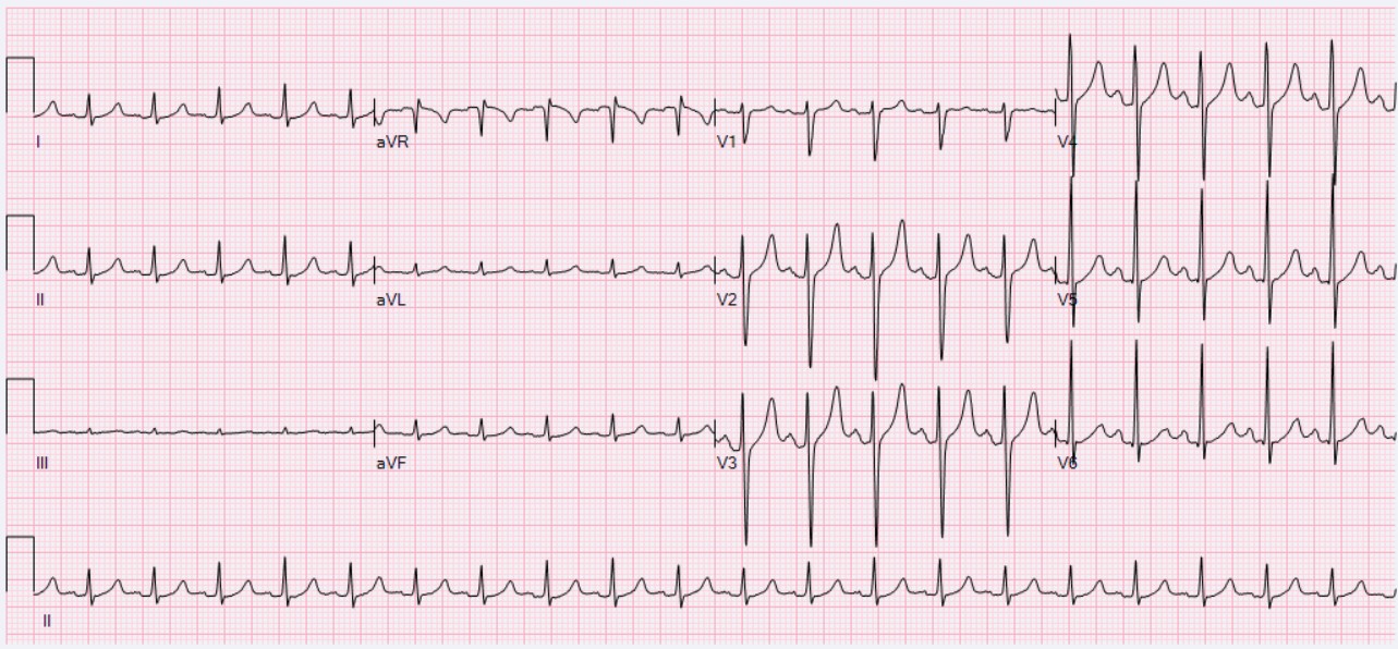
ECG: Sinus Tachycardia. - XR
-
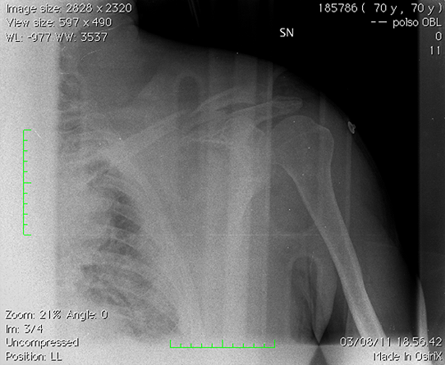
XR shows left midshaft clavicular fracture.
No pneumothorax was present on XR. - Further imaging: US / Angiogram
-
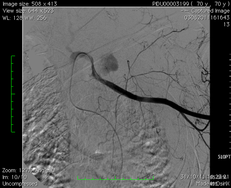
left subclavian arterial bleeding, 3 cm after homolateral vertebral artery. - CT
-
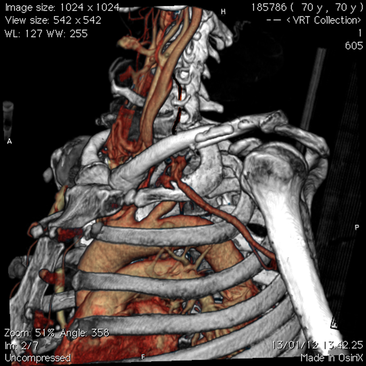
CT shows left subclavian arterial bleeding and the left midshaft clavicular fracture.
Last edited by a moderator: